43
FNAC nodules in the region of parotid, not necessarily within it.
Angiosarcoma
Different types of sarcoma are known to arise from blood vessels as angiosarcoma and Kaposi sarcoma. Angiosarcoma is a relatively rare target for FNA. The histological pattern of angiosarcoma is
variable, with the more differentiated tumours composed of vascular channels lined by tumour cells showing variable pleomorphism and atypia. Papillary extensions and cell rufis are rather common.
Obvious vessel-like structures are often difficult to find in poorly-differentiated angiosarcoma and tumour cells with paracentral nuclei and cytoplasmic vacuoles with single or small groups of
erythrocytes may be the only sign to indicate a vascular origin. In a subset of angiosarcoma, the epithelioid angiosarcoma has tumour cells with large and epithelial type of shape. Most angiosarcoma
stain for CD31 , with other useful markers being CD34.About 50% of epithelioid angiosarcoma stain for cytokeratins.
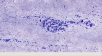
The cytological features of angiosarcoma in FNA material is very variable corresponding to those in histological sections. Atypical spindle cells, rounded and polygonal cells featuring variable
pleomorphism and atypia are common. Less frequently vacuolated atypical cells with erythrocytes in the vacuoles are found. Important pitfalls are spindle cell sarcoma and pleomorphic sarcoma of other
lines of differentiation, metastatic carcinoma and melanoma. Most often the cell population of angiosarcoma is diagnosed as malignant in smears. A specific diagnosis, however, is very difiicult or
impossible to render based on routinely stained smears and requires immunocytochemical stainings.
